Home » Without Label » Back Muscles Diagram - Human Muscle System Functions Diagram Facts Britannica - We think this is the most useful anatomy picture that you need.
Back Muscles Diagram - Human Muscle System Functions Diagram Facts Britannica - We think this is the most useful anatomy picture that you need.
Back Muscles Diagram - Human Muscle System Functions Diagram Facts Britannica - We think this is the most useful anatomy picture that you need.. The extensor muscles are attached to back of the spine and enable standing and lifting objects. When back development is the goal, stick to one of these variations. Likewise, there are muscles in other parts of the body that help support and move the spine. To learn more about the anatomy of the spine, watch this video. Muscles of lower back diagram in this image, you will find an occipital bone, sternocleidomastoid, trapezius, deltoid in muscles of the lower back diagram.
To learn more about the anatomy of the spine, watch this video. Nerves in your lower back. The extensor muscles are attached to back of the spine and enable standing and lifting objects. Three types of back muscles that help the spine function are extensors, flexors and obliques. The deltoid, teres major, teres minor, infraspinatus, supraspinatus (not shown) and subscapularis muscles (not shown) all extend from the scapula to the humerus and act on the shoulder joint.
Handcuff Muscles Brooklyn Reflexology from abcdefgh25.files.wordpress.com The deltoid, teres major, teres minor, infraspinatus, supraspinatus (not shown) and subscapularis muscles (not shown) all extend from the scapula to the humerus and act on the shoulder joint. Both the deltoid and the trapezius are firmly attached to the spine of the scapula. They span between the transverse and spinous processes of the regional vertebrae. Learn vocabulary, terms, and more with flashcards, games, and other study tools. Muscles of lower back diagram in this image, you will find an occipital bone, sternocleidomastoid, trapezius, deltoid in muscles of the lower back diagram. Back muscles, back muscle diagram. Curl your spine down, engaging your abdominal muscles, keeping your legs. The latissimus dorsi, also known as the lats or wings, are.
Names and diagram daniel nelson on january 1, 2019 2 comments !
The latissimus dorsi, also known as the lats or wings, are. These structures work together to support the body, enable a range of movements, and send messages from the. The semispinalis muscle, which is topographically divided into the semispinalis capitis , semispinalis cervicis and semispinalis thoracis . It is opposite from the chest, and the vertebral column runs down the back. The following diagram below is the diagrams of back muscle. The most common type of back pain is muscle pain—also called muscle strain or soft tissue strain. Exhale as you carefully bend forward from the waist. The muscles of the lower back help stabilize, rotate, flex, and extend the spinal column, which is a bony tower of 24 vertebrae that gives the body structure and houses the spinal cord.the spinal. For example, some muscles located in the chest also help move the shoulders. Below you'll see diagrams along with the names of the back muscles that may be the cause of your pain. They originate from the vertebrae and insert into the scapulae. Muscles of lower back diagram in this image, you will find an occipital bone, sternocleidomastoid, trapezius, deltoid in muscles of the lower back diagram. These muscles include the large paired muscles in the lower back, called erector spinae, which help hold up the spine, and gluteal muscles.
Exhale as you carefully bend forward from the waist. The deltoid, teres major, teres minor, infraspinatus, supraspinatus (not shown) and subscapularis muscles (not shown) all extend from the scapula to the humerus and act on the shoulder joint. This is a diagram of the larger and more surface muscles of the low back. Muscle anatomy games 12 photos of the muscle anatomy games anatomy muscle labeling games, anatomy muscle matching games, anatomy muscle naming game, muscle anatomy learning games, muscle anatomy memory game, human muscles, anatomy muscle labeling games, anatomy muscle matching games, anatomy muscle naming. Three types of back muscles that help the spine function are extensors, flexors and obliques.
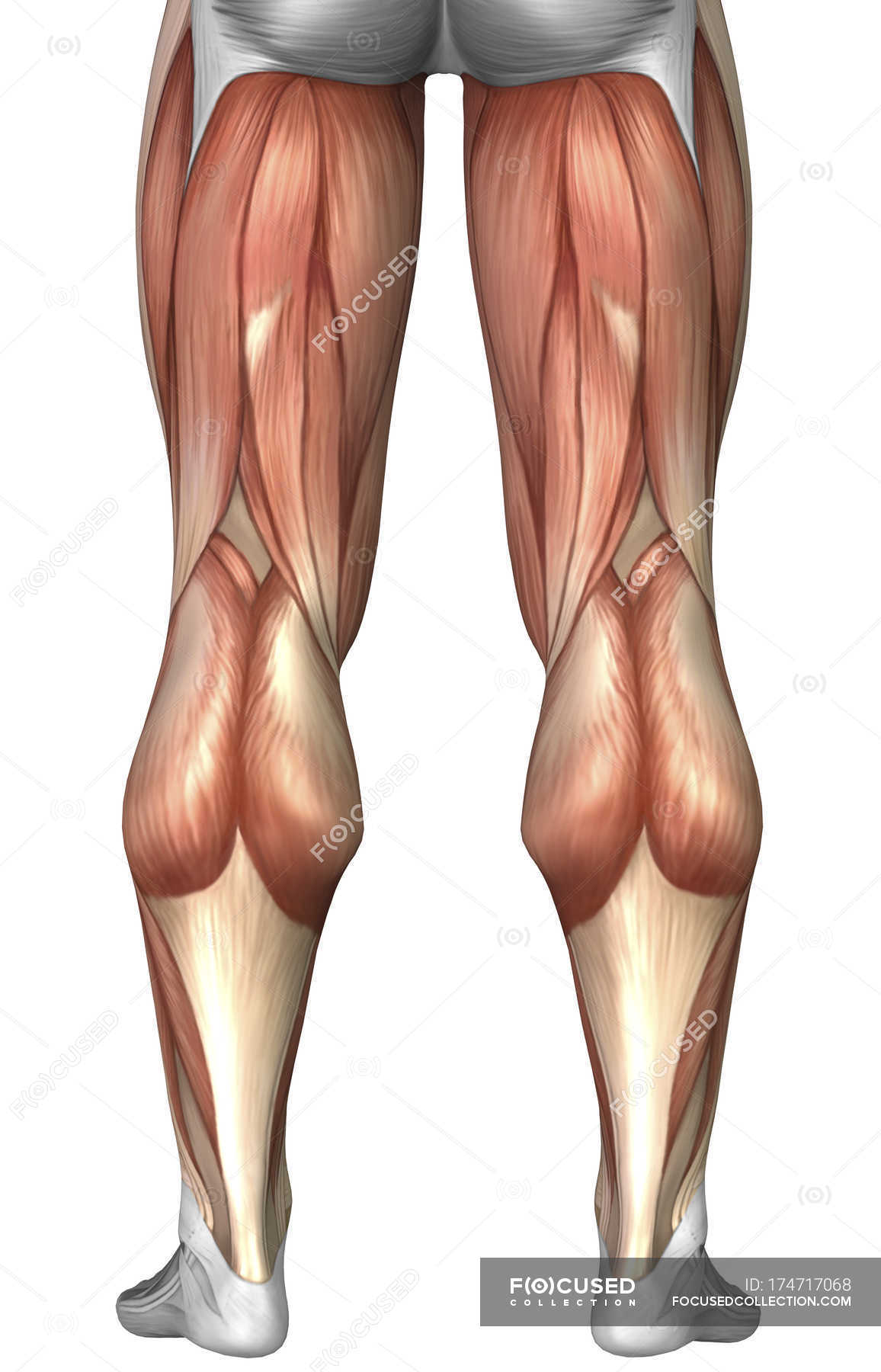
Diagram Illustrating Muscle Groups On Back Of Human Legs Vastus Lateralis Gracilis Stock Photo 174717068 from st.focusedcollection.com The back anatomy includes the latissimus dorsi, trapezius, erector spinae, rhomboid, and the teres major. Most of the time, back muscle pain is diagnosed then treated with little more than a prescription of rest, painkillers and muscle relaxants. The muscles of the lower back help stabilize, rotate, flex, and extend the spinal column, which is a bony tower of 24 vertebrae that gives the body structure and houses the spinal cord.the spinal. The part of the nerve that emerges out of the spine is called the nerve root. Symptoms of muscle pain include: It is opposite from the chest, and the vertebral column runs down the back. Creatine is now proving to be one of the most potent muscle growth accelerators giving excellent muscle mass increase and phenomenal strength increases order yours today. They originate from the vertebrae and insert into the scapulae.
Muscles of the back diagram.
They span between the transverse and spinous processes of the regional vertebrae. Superficial back muscles, intermediate back muscles and intrinsic back muscles.the intrinsic muscles are named as such because their embryological development begins in the back, oppose to the superficial and intermediate back muscles which develop elsewhere and are therefore classed as extrinsic muscles. The latissimus dorsi, also known as the lats or wings, are. What is the origin and insertion of the rhomboid minor and major muscle? Crane washes its wing tips. The deltoid, teres major, teres minor, infraspinatus, supraspinatus (not shown) and subscapularis muscles (not shown) all extend from the scapula to the humerus and act on the shoulder joint. The extensor muscles are attached to back of the spine and enable standing and lifting objects. It is opposite from the chest, and the vertebral column runs down the back. This picture also contains humerus, olecranon process of ulna, deep to tendon and so on. These muscles include the large paired muscles in the lower back, called erector spinae, which help hold up the spine, and gluteal muscles. Both the deltoid and the trapezius are firmly attached to the spine of the scapula. Names and diagram daniel nelson on january 1, 2019 2 comments ! This muscle is a major generator of lower back and hip pain, as well as being responsible for complaints of a burning sensation along the posterior superior iliac spine (psis) and sacroiliac joint.
Creatine research more than a sports supplement read more…. There are several different layers of muscles in your back that are often pulling in different and various directions. Lower back muscle diagram anatomy does degenerative disc disease affect the lower back muscle? When back development is the goal, stick to one of these variations. These structures work together to support the body, enable a range of movements, and send messages from the.
Muscles Of The Neck And Torso Classic Human Anatomy In Motion The Artist S Guide To The Dynamics Of Figure Drawing from doctorlib.info They span between the transverse and spinous processes of the regional vertebrae. Below you'll see diagrams along with the names of the back muscles that may be the cause of your pain. The human back extends from the buttocks to the posterior portion of the neck and shoulders. Others, like sumo deadlifts, have been shown in emg studies—and in the trenches—to focus more on other muscle groups than the back. These structures work together to support the body, enable a range of movements, and send messages from the. We hope this picture anatomy of back muscles diagram can help you study and research. The part of the nerve that emerges out of the spine is called the nerve root. Five pairs of lumbar spinal nerves labeled l1 to l5 branch off your spinal cord and exit through small holes between the vertebrae.
Superficial back muscles, intermediate back muscles and intrinsic back muscles.the intrinsic muscles are named as such because their embryological development begins in the back, oppose to the superficial and intermediate back muscles which develop elsewhere and are therefore classed as extrinsic muscles.
The transversospinal muscles gather three groups of back muscles: The following diagram below is the diagrams of back muscle. Creatine research more than a sports supplement read more…. They originate from the vertebrae and insert into the scapulae. The rhomboid muscle is activated as you bring and squeeze your scapula or shoulder blades back and together. This muscle is a major generator of lower back and hip pain, as well as being responsible for complaints of a burning sensation along the posterior superior iliac spine (psis) and sacroiliac joint. Creatine is now proving to be one of the most potent muscle growth accelerators giving excellent muscle mass increase and phenomenal strength increases order yours today. The muscles of the back can be arranged into 3 categories based on their location: This muscle is located on the upper portion of the back anatomy, underneath the trapezius. Muscles of the back diagram. Another common cause of lower back and hip pain is disc injury. As you can see, there are also have a spine of scapula deltoid, triceps brachii, latissimus dorsi. To learn more about the anatomy of the spine, watch this video.